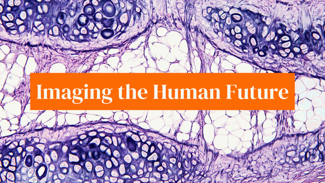
Imaging the Human Future: Breakthroughs in Medical Science
Introduction
The human body, a marvel of intricate complexity, orchestrates life’s dance through organs, tissues, cells, and proteins. While biomedical research offers glimpses into this complexity, it merely captures a moment in time.

The game-changer? Imaging technology. This powerhouse has the potential to provide real-time insights into biological processes, reshaping our understanding of human health and diseases.
At the forefront of this imaging revolution is the Chan Zuckerberg Initiative’s (CZI) Imaging Program.
Established to democratize access to imaging training and technology, the program funds cutting-edge technologies, delving deep into the human body for groundbreaking discoveries in curing, preventing, or managing diseases.
Integral to CZI’s work is the connection of experts, the creation of knowledge networks, and the development of tools and technologies.
In 2021, nearly 80 new grantees joined the initiative, contributing to the growth of the imaging tech tool Napari and advancing the visualization of proteins.
This collaborative effort aims to foster international networks of imaging experts, driving progress in the field.
As part of CZI’s 10-year science vision, an advanced imaging institute is in the works. This institute aims to propel revolutionary imaging technologies through high-risk, high-reward projects, marking a significant step toward transforming medical imaging.
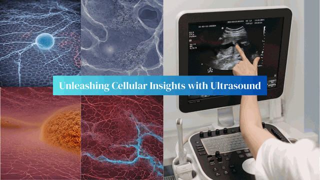
Mikhail Shapiro, an imaging grantee, is on the cusp of transforming ultrasound imaging. Currently confined to larger structures, ultrasound will soon delve into cellular functions deep within the body, courtesy of acoustic biomolecules.
This innovation opens avenues for understanding and treating various health issues, from neuroscience to gene therapy.
Breakthroughs in imaging science have birthed a dazzling light show revealing how neurons fire in a mouse brain.
Paul Hernandez-Herrera employs artificial intelligence to analyze vast amounts of data, creating a reliable and automated method to decipher the complexities of neuronal communication.
Delving into the world of trillions of cells and millions of proteins, Xiaoyu Shi and Jennifer Prescher are crafting methods in super-resolution microscopy.
Their collaboration aims to render 3D visualizations, making it easier to observe protein interactions at high resolution and understand their roles in health and disease.
Holger Müller and his team employ cutting-edge physics to maximize information retrieval from an electron beam, pushing quantum imaging to new heights.
Their work provides a behind-the-scenes look at the incredible technology employed for high-resolution imaging.

Imaging researcher Allison Dennis focuses on developing tools that combine light and ultrasound to screen for health issues during pregnancy, particularly preeclampsia.
By creating biocompatible nanoparticles as contrast agents, Dennis aims to enable imaging at greater depths, offering crucial insights for early diagnosis and treatment.
Scientist Ryan Cabeen merges science and art in vibrant 3D visualizations of a rat brain. His team’s work accelerates discovery in basic neuroscience by building the next generation of computational tools, making imaging research more accessible.
Rossana Melo employs high-resolution imaging to observe how immune cells respond to infections, including SARS-CoV-2. Her work not only enhances our understanding of infections but also strives to expand access to electron microscopy technologies in Brazil.
Scientist Prisca Liberali envisions the future by building open-source tools for imaging analysis. Her team, using the community-built napari tool, aims to create a flexible and easy-to-use framework for interactive plotting tools, fostering collaborative innovation.
Conclusion
In the realm of imaging science, these pioneers are pushing the boundaries of what we can see and understand about the human body.
The CZI Imaging Program’s commitment to fostering collaboration and innovation ensures that we are on the brink of a new era in healthcare, where imaging plays a central role in unraveling the enigmas surrounding human health and illness.
Frequently Asked Questions
How does acoustic biomolecule technology enhance ultrasound imaging?
Acoustic biomolecules enhance ultrasound imaging by allowing visualization of cellular functions deep inside the body.
They mark a paradigm shift, enabling researchers to observe specific functions of cells and molecules, not just larger structures.
Paul Hernandez-Herrera utilizes artificial intelligence to analyze vast amounts of data generated by imaging patterns of neuronal firing.
This technology aids in a reliable and automated understanding of the complexities of neuronal communication.
How do Xiaoyu Shi and Jennifer Prescher contribute to super-resolution microscopy?
Xiaoyu Shi and Jennifer Prescher are crafting methods in super-resolution microscopy to render 3D visualizations of protein interactions.
Their collaborative work aims to provide high-resolution insights into how millions of microscopic proteins in our cells function in health and disease.
Allison Dennis’s work involves developing biocompatible nanoparticles as photoacoustic contrast agents for imaging during pregnancy.
This innovation aims to enable imaging at significantly greater depths, offering crucial insights for the early diagnosis and treatment of complications like preeclampsia.
How does Prisca Liberali contribute to the future of imaging analysis?
Prisca Liberali is crafting open-source tools for imaging analysis using the community-built Napari tool. Her vision is to create a flexible and easy-to-use framework for interactive plotting tools, fostering collaborative innovation in the field of imaging science.
Get Access Now: https://bit.ly/J_Umma